Research Interests
Our lab is interested in examining the functional anatomy of attention and information selection in the healthy human brain, and how these neural circuits breakdown in schizophrenia and other neuropsychiatric and neurodevelopmental disorders. Our studies utilize multimodal neuroimaging techniques, including functional magnetic resonance imaging, scalp-recorded event-related potentials and behavioral neurocognitive testing batteries.
Facilities
ERP Facilities – Acquisiton and Analysis
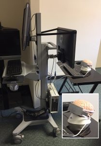
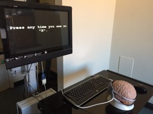
Located in the UNC Medical School Wings, the Electrophysiology Lab houses a fully equipped sound proof chamber to measure human ERP. The lab uses a 32-channel EEG system connected to Neuroscan software which acquires and analyzes ERP data. Video monitoring and intercom systems are located outside the chamber to track patients’ progress. An infrared eye-tracker and motion detection system also allow patient monitoring.
For more detailed information, click here.
MRI Acquisition
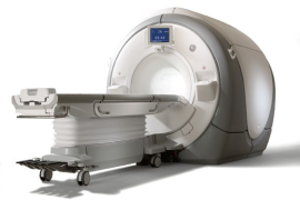 Some MRI scans are done at the Brain Imaging and Analysis Center (BIAC) in the Duke University Medical Center. The BIAC facilities include two General Electric MR750 3.0-Tesla research dedicated MR scanners, as well as a 7.0-Tesla Bruker research scanner used mostly for animal models. The BIAC also gives us access to MRI simulation facilites that will allow subjects to get used to the scanner environment while performing any tasks they are asked to do. For more information, click here.
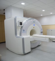 Other MRI scans are done at the Biomedical Research Imaging Center (BRIC) on the UNC-Chapel Hill campus. The BRIC facilities include a Siemens Magnetom Prisma 3T whole body scanner MRI scanner, a Siemens Biograph mMR 3TMR/PET scanner, a Siemens Magnetom 7T whole body MR scanner, as well as a Siemens Biograph mCT that combines high quality PET and CT imaging. Like the BIAC, the BRIC also has animal imaging and MRI simulation facilites. For more information, click here.
MRI Analysis
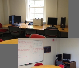 Access to multiple workstations (Windows and 64-bit Linux OS) is available at both the UNC-CH and Duke (BIAC) sites. In addition to these workstations, we have access to the Neuroimaging Lab of the NDRC workstations. These facilities exist to serve funded neuroimaging projects for approved NDRC investigators. All projects utlize the processing technology for quantitative measurements of structural and functional MRI. In addition, the Neuro Image Research and Analysis Lab of UNC-CH is available for assistance with the development and utilization of advanced image analysis techniques.
fNIR Acquisition and Analysis
 The NIRL also has a Biopac fNIR Imager Model 1100, a non-invasive oxygenation and blood volume trend imager. The fNIR Imager is designed to allow one to track relative oxygen consumption as well as changes in blood volume.

